
Tib post tendinopathy: symptoms, causes, and treatment
- 3 hours ago
- 9 min read
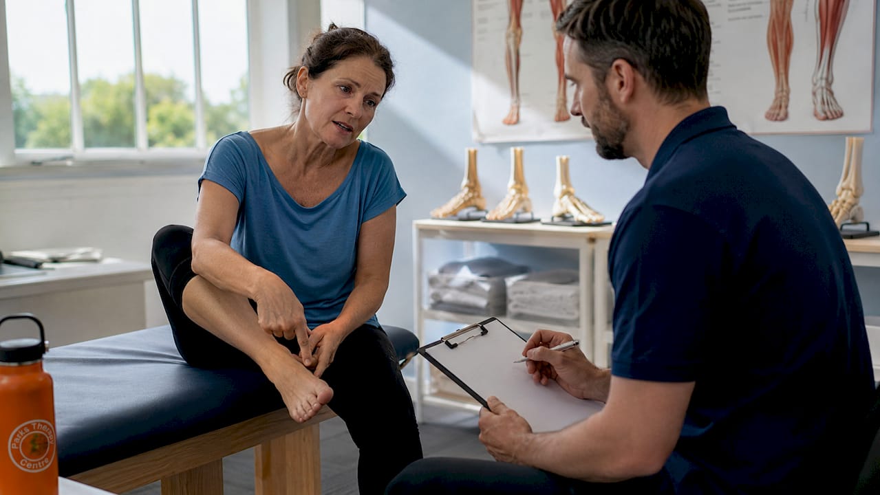
What is posterior tibialis tendinopathy?
Posterior tibialis tendinopathy, often referred to as tib post tendinopathy, is a condition involving injury or degeneration of the tibialis posterior tendon. This tendon runs along the inside of your ankle and plays a central role in supporting the arch of your foot during weight-bearing activity. When it becomes overloaded or damaged, pain and reduced foot function follow.
The condition progresses through distinct stages. It begins as tendonitis, an acute inflammatory response, and can advance to tendinosis, where the tendon undergoes chronic structural degeneration without true inflammation. Left unmanaged, this progression to PTTD (posterior tibial tendon dysfunction) leads to arch collapse and significant loss of function.
Key facts about the condition at a glance:
The tibialis posterior tendon is the primary dynamic stabiliser of the medial arch
Tendonitis refers to acute inflammation; tendinosis describes chronic, degenerative change
Posterior tibial tendon dysfunction (PTTD) is the advanced stage, now also termed progressive collapsing foot deformity (PCFD)
Pain typically appears along the inside of the ankle, the instep, and the lower leg
Most patients present with one affected foot rather than both
What are the symptoms of tib post tendinopathy?
Pain along the inside of the ankle is the hallmark symptom, and it tends to be most noticeable during or after activity. Many patients also report morning stiffness that eases once they begin moving, only to worsen again after prolonged standing or walking.
Recognising the full range of symptoms helps you seek care before the condition advances:
Pain and tenderness along the inner ankle and lower leg, particularly on direct pressure
Difficulty or inability to perform a single-leg heel raise on the affected side
Mild to moderate weakness when rising onto the toes
Swelling along the course of the tendon, from behind the inner ankle bone down to the arch
Pain that worsens with running, jumping, or extended walking
In later stages, a visible flattening of the foot arch and forefoot drift outward
One clinical sign of advanced arch collapse is the ‘too many toes’ sign: when viewed from behind, additional toes become visible on the outer side of the foot, indicating that the forefoot has abducted due to tendon failure. Patients who delay care often reach this stage without realising how far the condition has progressed.
How common is tibialis posterior tendinopathy?
Prevalence: Posterior tibialis tendinopathy affects between 3.3% and 10% of the population, with the highest rates seen in women aged 40–50 years. The condition almost always affects one foot rather than both.
Middle-aged women represent the group most frequently affected, though the condition does occur across a broader age range, particularly in people with relevant risk factors such as obesity or diabetes. The unilateral presentation is notable: bilateral involvement is uncommon and, when present, usually points to a systemic inflammatory condition rather than mechanical overload alone.
The gap between how common the condition is and how often it is correctly identified early remains a concern. Early-stage tibialis posterior pain is frequently misinterpreted as a general ankle sprain or flat-foot discomfort, which delays targeted treatment and allows structural changes to develop.
What causes posterior tibialis tendinopathy?
Repetitive stress and microinjury to the tendon are the primary drivers. When activity levels increase too quickly, the tendon sustains small injuries faster than the body can repair them, gradually weakening the structure. This is why a sudden spike in running mileage or prolonged standing at work often precedes the onset of symptoms.
Several factors raise the risk of developing a posterior tibial tendon injury:
Age: Tendon tissue becomes less resilient with age, particularly from the mid-forties onward
Excess body weight: Increases the load through the tendon with every step
Diabetes: Linked to collagen disorganisation and reduced tendon healing capacity
Inflammatory conditions: Rheumatoid arthritis and psoriatic arthritis accelerate tendon degeneration
Overpronation and flat feet: Alter load distribution and place the tendon under sustained stretch
Unsupportive footwear: Sandals and flat, flexible-soled shoes remove the structural support the tendon needs
Prolonged standing or walking: Particularly with a sudden increase in duration or intensity
Corticosteroid use: Prolonged oral corticosteroids and certain antibiotics (fluoroquinolones) weaken tendon tissue
Previous foot or ankle injury or surgery: Creates altered mechanics that stress the tendon
Smoking: Impairs tissue perfusion and slows healing
High-impact sports: Especially those involving repetitive jumping or rapid direction changes
Biomechanical contributors deserve particular attention. Overpronation places the tibialis posterior under continuous eccentric load, and patients with pre-existing flat feet carry a structurally elevated baseline risk.
Treatment options for tibialis posterior tendinopathy
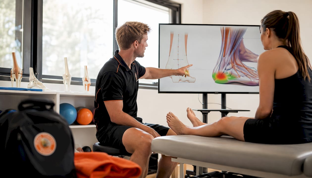
The good news is that most patients improve with conservative management, though tendons heal slowly and recovery typically takes several months. The goal is not rest in isolation but rather optimal load management: reducing aggravating activity while maintaining enough load to stimulate tendon adaptation.
Conservative treatment components:
Footwear modification: Switch to structured running trainers or hiking boots with a slight heel elevation. Flat, flexible-soled shoes increase tendon stress and should be avoided
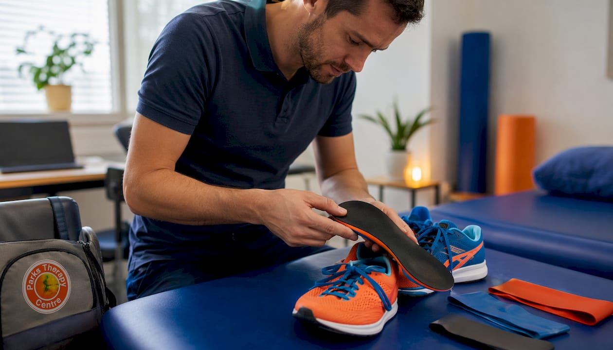
Orthotics: Custom or prefabricated insoles with medial arch support reduce the load on the tendon during weight bearing. University of California Biomechanics Laboratory (UCBL) orthoses are used in more advanced cases
Bracing: An ankle-foot orthosis (AFO) or supportive brace can offload the tendon significantly, particularly during flare-ups. Guidance on supportive bracing options can help you understand which device suits your stage of recovery
Physical therapy: A structured physiotherapy programme targeting tibialis posterior strengthening is the cornerstone of recovery
Activity modification: Reduce high-impact loading while maintaining low-impact movement such as swimming or cycling
Analgesics: Paracetamol or short-term NSAIDs can help manage pain, but anti-inflammatory medication should not be relied upon long-term as tendinosis involves degeneration rather than active inflammation
Pro Tip: Switch to structured trainers with a slight heel and lace them firmly from the toe upward. Properly laced, supportive footwear reduces tendon stress during every step and is one of the most immediately effective changes you can make.
Surgery is reserved for patients who have not responded to several months of conservative care, or those with an acute tendon tear. Procedures range from tenosynovectomy in early cases to medial calcaneal osteotomy with tendon repair, and in advanced PTTD, surgical reconstruction or arthrodesis may be required. Patients must avoid glucocorticoid injections directly into the tendon sheath, as these accelerate degeneration and raise the risk of rupture.
Exercise guidance for tibialis posterior tendinopathy recovery
Complete rest is counterproductive for tendon healing. The tendon needs controlled load to stimulate collagen remodelling and regain strength. The principle of optimal loading means you exercise within a tolerable pain range, typically no more than 3–4 out of 10 on a pain scale, and monitor how the tendon responds in the 24 hours after each session.
Targeted exercises for recovery include:
Seated calf raises (bilateral): Begin with both feet, progressing to single-leg as strength improves. This loads the tibialis posterior in a controlled, low-impact way
Single-leg heel raises: The key functional test and a core rehabilitation exercise. Start with support from a wall or chair and build toward unassisted repetitions
Tibialis posterior resisted inversion: Using a resistance band, invert and plantarflex the foot against resistance to directly strengthen the tendon
Towel scrunches and toe spreads: Improve intrinsic foot muscle strength, which supports arch mechanics
Calf stretching (gastrocnemius and soleus): Tight calf muscles increase tendon load; regular stretching reduces this
Balance and proprioception work: Single-leg standing on a wobble board or foam surface restores neuromuscular control around the ankle
Progression should be gradual. If pain increases beyond a tolerable level during or after exercise, reduce the load rather than stopping entirely. A physiotherapist can design a structured home exercise plan that matches your current stage of recovery and adjusts as you improve.
How to manage pain flare-ups during recovery
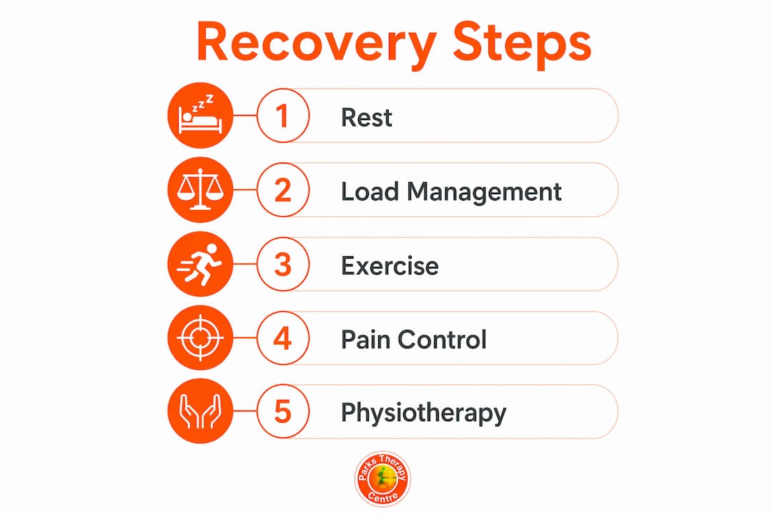
Flare-ups are a normal part of tendon rehabilitation and do not mean the condition is worsening permanently. They most commonly occur after a sudden increase in activity, a change in footwear, or a period of prolonged standing.
Practical steps to manage a flare-up:
Reduce load temporarily: Cut back on the activity that triggered the increase in pain, but avoid complete rest
Apply ice: A cold pack wrapped in a cloth applied to the inner ankle for 15–20 minutes can reduce local discomfort
Review footwear: Revert to your most supportive trainers and check that laces are properly fastened
Use analgesics appropriately: Paracetamol for short-term pain relief; NSAIDs for brief periods if tolerated and not contraindicated
Modify exercise: Temporarily reduce resistance or repetitions rather than stopping the rehabilitation programme altogether
Elevate the foot: Particularly useful if swelling accompanies the flare-up
Seek professional advice if pain persists beyond a few days despite these measures, if swelling increases markedly, or if you notice a sudden change in foot shape. Recurrent flare-ups that do not settle with self-management are a clear signal that a physiotherapy review is needed.
Why expert physiotherapy care makes a difference for tibialis posterior tendinopathy
A physiotherapist does more than prescribe exercises. They assess the full picture: your foot mechanics, muscle strength, gait pattern, footwear, and activity demands. From that assessment, they build an individualised rehabilitation plan that matches your specific stage of tendon damage and your recovery goals.
The benefits of professional physiotherapy care include:
Accurate diagnosis: Distinguishing tibialis posterior tendinopathy from other causes of medial ankle pain, including tarsal tunnel syndrome and deltoid ligament injury
Graded loading programmes: Structured to stimulate tendon healing without aggravating symptoms
Gait and biomechanical correction: Identifying and addressing overpronation, leg length discrepancy, or altered walking patterns that perpetuate tendon stress
Orthotic prescription and review: Ensuring insoles and bracing are correctly fitted and appropriate for the stage of the condition
Education and self-management: Equipping patients with the knowledge to manage flare-ups and progress safely at home
NHS guidance supports physiotherapy as the primary intervention for tibialis posterior tendinopathy, particularly when self-managed conservative measures have not produced improvement within 6–12 weeks. Physical therapy focusing on strengthening and optimal load consistently achieves better long-term functional outcomes than rest alone.
Parkstherapycentre has provided expert musculoskeletal physiotherapy since 1986, with clinics across Bedfordshire and Buckinghamshire. The multidisciplinary team includes physiotherapists, podiatrists, and sports injury specialists who work together to deliver patient-centred care for conditions including tibialis posterior tendinopathy. Patients with more complex presentations, including those who have undergone surgery, benefit from the centre’s expertise in post-surgical ankle rehabilitation.
How is tibialis posterior tendinopathy diagnosed?
Diagnosis begins with a clinical examination. A physiotherapist or clinician will assess tenderness along the tendon, test single-leg heel raise capacity, and look for signs of arch collapse. Tenderness is typically maximal when the foot is held in inversion and plantarflexion, a position that places the tendon under direct palpation stress.
The single-leg heel raise test is particularly informative. An inability to rise fully onto the toes on the affected side, or pain that prevents the attempt, points strongly toward tibialis posterior involvement. The ‘too many toes’ sign, assessed from behind the standing patient, indicates forefoot abduction and advanced tendon failure.
When clinical findings are inconclusive or symptoms persist despite initial treatment, imaging provides additional detail. MRI and ultrasound can confirm tenosynovitis, map the extent of tendinosis, and identify partial or complete tears. MRI offers superior soft-tissue resolution for complex cases, while ultrasound allows dynamic assessment of tendon movement. Plain X-rays add little for tendon pathology but may be used to assess bony alignment in advanced PTTD.
What happens if tibialis posterior tendinopathy is left untreated?
Without treatment, the condition follows a predictable and worsening course. The tendon progressively weakens, surrounding ligaments come under increasing strain, and the medial arch gradually collapses. This is the pathway to posterior tibial tendon dysfunction and, ultimately, adult-acquired flatfoot deformity.
Functional consequences become significant as the condition advances. Walking distances shorten, stair climbing becomes painful, and activities that were previously unremarkable, such as standing at a kitchen counter, cause discomfort. In severe cases, bony deformity develops in the hindfoot and midfoot, at which point conservative treatment is no longer sufficient and surgical reconstruction becomes necessary. Early detection and consistent rehabilitation are the most effective ways to prevent this trajectory.
Conditions that can mimic tibialis posterior tendinopathy
Several conditions produce medial ankle and foot pain that closely resembles tibialis posterior tendinopathy, and accurate differentiation matters because treatment approaches differ considerably.
Tarsal tunnel syndrome involves compression of the tibial nerve as it passes through the tarsal tunnel behind the medial malleolus. Patients typically describe burning, tingling, or numbness rather than the aching, activity-related pain of tendinopathy. Tinel’s sign (tapping over the tarsal tunnel to reproduce symptoms) helps distinguish it clinically.
Deltoid ligament sprain or insufficiency produces medial ankle pain and may coexist with tibialis posterior pathology in advanced PTTD. Tenderness is anterior to the medial malleolus rather than along the tendon course, and MRI can confirm ligament involvement.
Flexor hallucis longus tendinopathy causes pain behind the medial ankle, but symptoms are more localised to the big toe flexor and worsen specifically with toe push-off rather than arch loading.
Spring ligament injury is increasingly recognised as a cause of medial arch pain and flatfoot deformity, sometimes in the absence of tibialis posterior tendon damage. Patients present with normal inversion strength but tenderness anterior to the medial malleolus, and MRI confirms the diagnosis.
Stress fracture of the navicular produces dorsal midfoot pain that worsens sharply with activity and eases completely with rest, a pattern distinct from tendinopathy. Point tenderness over the navicular and MRI or CT imaging confirm the diagnosis.
A thorough clinical assessment, combined with imaging where indicated, is the only reliable way to distinguish these conditions and direct the right treatment.
Key takeaways
Tib post tendinopathy responds well to early, structured rehabilitation centred on optimal load management, appropriate footwear, and targeted strengthening of the tibialis posterior muscle.
Point | Details |
Prevalence and risk groups | The condition affects 3.3%–10% of the population, most commonly women aged 40–50, usually in one foot only. |
Optimal loading over rest | Complete rest delays healing; controlled exercise within a tolerable pain range drives tendon recovery. |
Footwear is a first-line intervention | Structured trainers with a slight heel and proper lacing reduce tendon stress immediately and meaningfully. |
Early diagnosis prevents progression | Untreated tendinopathy advances to arch collapse and adult-acquired flatfoot, which may require surgery. |
Physiotherapy delivers better outcomes | A graded, individualised physiotherapy programme consistently outperforms self-managed rest for long-term function. |
Recommended