
Plantar plate tear treatment: a practical recovery pathway
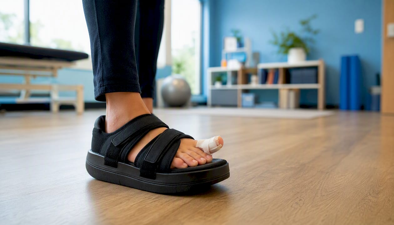
Most plantar plate tears respond to conservative mechanical stabilisation and structured rehabilitation, not surgery. Restricting barefoot walking, wearing a stiff-soled or rocker-bottom shoe, taping the affected toe into a plantarflexed position, and starting pain-limited strengthening work is the standard first move, and it works for the majority of people who catch the problem early.
Surgery is reserved for a smaller group: those with ongoing joint instability, a progressive crossover or floating toe deformity, or pain that fails to settle after a genuine trial of conservative care, usually several months.
Here’s what to do in the first few weeks:
Cut back on barefoot walking and swap flexible trainers for a rigid or rocker-soled shoe.
Tape the affected toe to hold it in a slightly plantarflexed position, reducing strain on the healing plate.
Apply ice for short periods if the joint is acutely swollen or hot.
Begin gentle, pain-limited toe exercises rather than resting completely.
Book an assessment if pain, swelling or toe drift hasn’t improved within 6 to 12 weeks, or sooner if you notice sudden deformity or the toe visibly floats above the ground.
Conservative care tends to suit people picked up early, with mild to moderate instability and no fixed deformity. Surgery becomes the realistic option for those with a chronic tear, visible toe crossover, or a joint that keeps subluxing despite consistent taping and strengthening.
Key takeaways
Most plantar plate tears heal with mechanical offloading, taping, and staged rehabilitation over 3 to 6 months, with surgery reserved for persistent instability or progressive deformity.
Point | Details |
Start conservative | Stiff or rocker-soled footwear, taping, and pain-limited exercise are the appropriate first step for nearly everyone. |
Give it time | Ligament healing genuinely takes 3 to 6 months; judge conservative treatment against that window, not a fortnight. |
Watch for red flags | Sudden deformity, floating toe, or numbness warrant a faster assessment than routine forefoot pain. |
Surgery has a clear role | It’s indicated for persistent instability, progressive crossover deformity, or failed conservative care, with pooled outcomes showing strong pain and function gains. |
Get assessed early | Parkstherapycentre’s podiatry and physiotherapy team can diagnose, start conservative treatment, and coordinate surgical referral or post-op rehab if needed. |
Table of Contents
What is a plantar plate tear and why does it happen?
The plantar plate is a tough, fibrocartilaginous structure that sits underneath each lesser metatarsophalangeal joint (the knuckle where your toes meet the ball of the foot). Think of it as a shock-absorbing hammock strung beneath the joint capsule, anchoring the base of the toe and resisting the upward force that push-off generates.
Every time you walk, run, or rise onto your toes, the plate stops the toe from hyperextending upward and keeps it tracking straight rather than drifting sideways. It’s a static stabiliser, doing quiet structural work rather than moving anything itself. When it frays or tears, that control disappears, and the toe can start to sit slightly elevated, drift towards its neighbour, or eventually cross over the toe beside it.
Injury tends to happen in one of three ways. The most common is chronic attrition: years of repetitive load gradually wearing down the fibres, much like a rope fraying rather than snapping. Less commonly, a single hyperextension injury, such as stubbing the toe hard or landing awkwardly from a jump, tears the plate acutely. Inflammatory joint disease, particularly rheumatoid arthritis, can also weaken the tissue directly, making tears more likely even without unusual mechanical stress. Across all these mechanisms, chronic attrition is the most frequently reported cause, which is part of why the injury often creeps up rather than announcing itself.
The second toe is affected more often than any other, largely because it frequently bears the longest metatarsal in the row, taking a disproportionate share of forefoot load with every step.
Who is most at risk of a plantar plate injury?
Certain foot shapes and habits load the second and third metatarsal heads more heavily than they’re built to withstand, and that mechanical overload is the thread running through most cases.
Mechanical risk factors:
A long second metatarsal relative to the first, shifting load onto that joint during push-off.
Tight calf muscles (gastrocnemius or soleus), which increase forefoot pressure by limiting ankle dorsiflexion.
Forefoot overload from a cavus (high-arched) foot type or a hypermobile first ray.
Footwear with a narrow toe box or unsupportive, overly flexible soles.
Systemic and activity-related risk factors:
Inflammatory arthropathies such as rheumatoid arthritis, which weaken periarticular soft tissue.
Sports and activities involving repetitive push-off, including running, dancing, and court sports.
Age-related changes in tissue elasticity, making tears more common from the late 40s onward.
Long-standing use of high heels, which shifts sustained load onto the forefoot.
Several of these factors are modifiable, and that matters more than it might seem. Swapping unsupportive trainers for a stiffer, well-fitted shoe, reducing running volume temporarily, and building intrinsic foot strength all lower the tensile strain on the plate during recovery. Ignore them, and even a well-executed rehab plan will keep getting undermined by the same forces that caused the tear in the first place.
What are the symptoms of a plantar plate tear?
The hallmark symptom is a focal, deep ache directly under the ball of the foot at the base of one toe, often described as feeling like walking on a stone or a persistent bruise that never quite fades. It typically worsens during the push-off phase of walking or running, when the toe is asked to extend and the plate is under greatest tension.
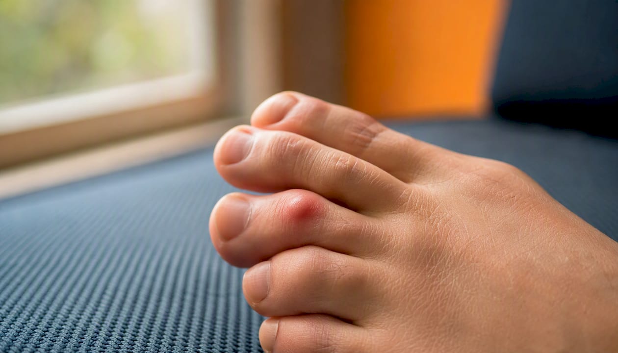
Swelling around the joint is common, and some people notice the toe starting to sit slightly higher than its neighbours, or drifting sideways with time. As the tear progresses, a hammer toe or crossover toe deformity can develop, where the affected toe angles over or under the toe beside it.
Clinicians rely on a handful of hands-on tests to confirm what imaging alone can’t always show. The most useful is the vertical stress test, sometimes called the drawer test: the clinician stabilises the metatarsal with one hand and applies upward pressure to the base of the toe with the other, checking how much the joint translates vertically compared with the unaffected side. Excess movement, or reproduction of your usual pain, points strongly towards a plantar plate tear. Reduced plantar grip, where the toe can’t actively press down against resistance, is another common finding, alongside early signs of hammer toe formation on close inspection.
A useful clinical marker: the drawer test is one of the most consistently cited physical examination findings for plantar plate instability, and it’s often the deciding factor in whether a clinician moves straight to imaging or manages the case conservatively based on exam findings alone.
Certain signs warrant faster attention rather than a “wait and see” approach. Sudden toe dislocation, a rapidly worsening deformity, numbness or colour change in the toe (suggesting neurovascular compromise), or signs of infection such as spreading redness and warmth should prompt an urgent assessment rather than a routine booking.
How is a plantar plate tear diagnosed?
Clinical examination remains the backbone of diagnosis, and for many straightforward presentations, imaging isn’t strictly necessary before starting conservative treatment. A clinician will typically combine your symptom history with palpation, the vertical stress test, and observation of toe alignment to reach a working diagnosis.
Imaging becomes more useful in specific scenarios: when conservative treatment hasn’t helped after a reasonable trial, when surgery is being planned, or when the presentation is atypical and other diagnoses need ruling out.
Weight-bearing X-rays show bony alignment, the metatarsal parabola (the relative lengths of the metatarsals), and any early arthritic change, but they don’t visualise the soft tissue plate itself.
Ultrasound allows dynamic, real-time visualisation of the plate under stress, and can pick up partial tears while the joint is moved through its range, though results depend heavily on operator experience.
MRI gives the clearest picture of tear extent and any associated pathology, such as synovitis or collateral ligament involvement, and is the modality most surgeons want before planning a repair.
Clinical and surgical grading systems, such as the one described by Coughlin, correlate what’s found on examination with the likely anatomical extent of the tear. That matters practically: grading helps predict whether a straightforward repair is feasible, or whether more extensive reconstruction, such as a tendon transfer, is likely to be needed.
Conservative treatment: what actually works first
The underlying principle of every non-surgical plantar plate injury treatment is the same: reduce the tensile strain running through the healing tissue while correcting whatever biomechanical factor pushed it past its limit in the first place. Get that balance right and most tears settle without ever needing a scalpel.
Footwear is the foundation. A stiff-soled shoe, ideally with a rocker profile built into the outsole, prevents the toe from having to bend through its full range during push-off. That single change removes a huge amount of load from the injured joint. Avoid high heels and anything with a soft, flexible forefoot, and resist the temptation to go barefoot around the house while the tear is healing, since bare feet offer zero mechanical protection.
Taping and strapping hold the toe in a slightly plantarflexed position, counteracting the upward drift that damages the plate further. This isn’t a permanent fix. Taping and offloading devices reduce motion and strain without correcting underlying bone alignment; they buy the tissue time to heal while any longer-term biomechanical issue is addressed separately. As a general guide, strapping continues for around 4 to 6 weeks, though your clinician will adjust that based on how the tear is progressing.
Orthoses and metatarsal pads work alongside taping rather than replacing it. A podiatrist typically prescribes a custom or semi-custom insole with a metatarsal dome placed just behind the painful joint, redistributing pressure away from the injured metatarsal head and onto the surrounding structures.
Pain relief is usually short-term and conservative: ice in the early, acutely inflamed phase, and NSAIDs used sparingly if there are no contraindications. One point clinicians are consistently firm about: steroid injections should not be given directly into the plantar plate, because corticosteroids can weaken the very ligament tissue you’re trying to protect, raising the risk of a partial tear progressing to a full rupture. For chronic cases (generally beyond three months) that haven’t responded to standard mechanical care, some clinicians consider PRP or amniotic injections as an adjunct, but these supplement mechanical stabilisation rather than substitute for it, as explained in peptides for tendon repair.
Set your expectations around weeks to months, not days. Ligament injuries in the foot typically take 3 to 6 months to heal fully, and rushing back into unsupportive footwear or high-impact activity before that window closes is the single most common reason conservative treatment fails.
Pro Tip: Buy your rocker-soled or stiff footwear before you need it, not after the pain has already flared. Breaking a new pair in gently while you’re symptomatic makes the whole recovery smoother, rather than adding a fresh irritation on top of an already sensitive joint.
Physiotherapy and rehabilitation: a phased approach
Rehabilitation for a plantar plate tear works best as a staged progression, gradually reintroducing load rather than jumping straight from rest to full activity.
Phase 1 (weeks 1 to 2): protect and calm things down. The priority here is pain control and protecting the healing tissue. Gentle isometric holds (pressing the toe down against light resistance and holding, without movement) and pain-limited range-of-motion work keep the joint from stiffening without stressing the plate.
Phase 2 (weeks 2 to 6): build intrinsic strength. This is where progressive intrinsic foot strengthening begins in earnest, alongside continued metatarsal pad use and taping. Towel scrunches, short foot exercises, and toe-spreading drills rebuild the small muscles that support the arch and the toe joints. Gait modifications also matter at this stage, particularly encouraging a shorter stride and pushing off through the big toe rather than the injured lesser toe.
Phase 3 (6+ weeks onward): reload and return. Progressive resisted toe strengthening (using resistance bands or manual resistance) and balance and proprioception work prepare the foot for higher demands. A graded return-to-run or return-to-walk programme follows, increasing distance and intensity in small increments and backing off if pain returns.
Escalate care and seek reassessment if strength doesn’t improve despite consistent rehab, if the deformity keeps progressing, or if you’re still functionally limited well past the expected timeline. Rehabilitation done well restores toe strength and reduces symptoms within roughly 4 to 6 weeks for many people, so a lack of progress by that point is worth flagging rather than pushing through.
Pro Tip: If you’re doing isometric toe presses at home, try pacing them to a metronome, roughly one press every two seconds. It sounds unnecessary, but rhythmic pacing during isometric work reduces the nervous system’s natural inhibition of the muscle, helping you build strength faster than pressing at random, inconsistent intervals.
When is surgery for a plantar plate tear considered?
Surgery moves from “possible” to “recommended” in a fairly specific set of circumstances, and it’s rarely the first conversation a clinician has with a new patient.
The clearest indications are persistent pain that hasn’t responded to a genuine conservative trial (typically several months of consistent taping, footwear change, and rehab), frank joint instability or dislocation that doesn’t reduce with conservative measures, and a progressive deformity such as a crossover toe that keeps worsening despite everything tried so far.
Before any operation, preoperative assessment usually includes MRI or ultrasound to confirm tear extent, weight-bearing X-rays to assess alignment and metatarsal length, and a detailed discussion about whether additional procedures, such as a Weil osteotomy or hammertoe correction, will be needed alongside the plate repair itself.
Honest counselling matters here. A good surgeon will walk you through realistic outcome expectations, the likely recovery timeline, and risks such as postoperative stiffness or a “floating toe” (where the toe sits elevated and doesn’t touch the ground properly afterwards). If the plate tissue itself turns out to be poor quality once the surgeon is inside the joint, the plan may shift towards a tendon transfer rather than a direct repair.
Surgical options and what the evidence shows
Several surgical approaches exist, and the right one depends on tear chronicity, deformity severity, tissue quality, and what you need your foot to do afterwards.
Dorsal direct repair is now the most commonly used technique and is often combined with a Weil osteotomy, a shortening cut in the metatarsal that improves access to the joint and corrects length imbalance at the same time. There’s a genuine trend in current practice towards this anatomic repair approach over older tenotomy techniques, largely because it preserves more of the joint’s natural function.
Plantar approach repair accesses the tear directly from underneath the foot rather than through the top of the joint, though it’s reported less frequently in the surgical literature than the dorsal route.
Flexor tendon transfer becomes the fallback when the plantar plate tissue itself is too degraded to repair directly, rerouting a flexor tendon to take over the stabilising job the plate can no longer do.
Metatarsal osteotomy (commonly the Weil osteotomy) is frequently performed alongside repair to shorten an overly long metatarsal and reduce the load driving the original injury.
A staged way of thinking about severity helps frame the decision: milder cases may need only realignment, moderate cases typically combine plantar plate repair with an osteotomy, and severe, chronic cases sometimes require tendon transfer or, rarely, resection of the metatarsal head.
The pooled evidence on outcomes is genuinely encouraging. A meta-analysis covering 12 studies and 537 repaired tears found a pooled mean pain reduction of around 5 points on the VAS scale, alongside an average AOFAS functional score improvement of roughly 40 points, with most follow-up data extending to mid-term periods. That’s a substantial functional gain by most surgical standards, though it’s worth being clear-eyed about what sits behind the number: the dorsal approach dominates this evidence base, and outcomes largely reflect repairs performed alongside a Weil osteotomy rather than isolated plate repair. Extrapolating those results to a dorsal repair done in isolation carries more uncertainty than the headline figures suggest.
Surgical risk | What it involves |
Joint stiffness | Reduced range of motion at the MTP joint after healing, sometimes needing extra mobilisation work |
Floating toe | The toe sits elevated and doesn’t make full contact with the ground during walking |
Infection | Standard surgical site risk, managed with wound care and monitoring |
Recurrence | Instability or pain returning, more likely with poor tissue quality or unaddressed biomechanics |
Transfer deformity | Excess load shifting to an adjacent toe or metatarsal after surgery |
The decision between these options ultimately rests on four things: how long the tear has been present, how severe the deformity has become, the quality of the remaining tissue once the surgeon can assess it directly, and what you personally need your foot to do, whether that’s running competitively again or simply walking without pain.
What does recovery after plantar plate surgery look like?
Recovery follows a fairly predictable arc, though exact timings vary by surgeon and technique.
Immediately after surgery, expect a period of immobilisation or protected weightbearing in a surgical shoe or boot, with attention to wound care and swelling control through elevation and Coban wrapping. Early loading is deliberately restricted here, since asking the repair to bear force too soon is one of the more common reasons for a suboptimal result.
Recovery stage | Typical timing | What’s happening |
Immediate post-op | Initial weeks | Protected weightbearing, wound care, swelling management, sutures typically removed around two weeks |
Early rehab | Weeks 2 to 6 | Gradual introduction of protected range-of-motion work, careful loading progression |
Later rehab | Weeks 6 to 12+ | Progressive strengthening, gait retraining, planning return to sport or work |
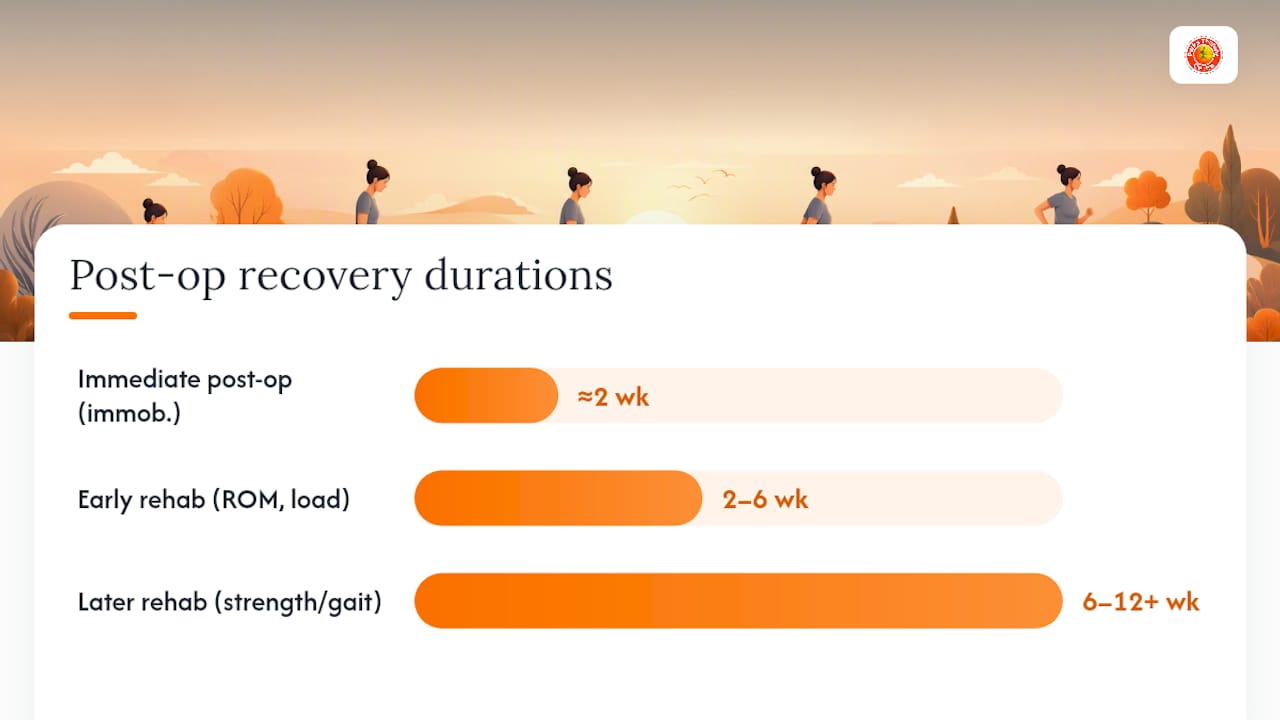
Throughout the early phases, avoiding early dorsal force on the toe is a key precaution, since pushing the toe upward too soon can strain the repair before it’s had time to mature. Range-of-motion work is introduced gradually and deliberately, balancing the risk of stiffness against the risk of overloading a repair that’s still healing. Return to sport or physically demanding work is judged against functional milestones (strength, pain-free range, gait quality) rather than a fixed calendar date, which is why timelines can vary noticeably between people who had similar surgery.
What happens if a plantar plate tear goes untreated?
Left alone, a plantar plate tear rarely stays the same. Without offloading or rehabilitation, the mechanical forces that caused the tear keep acting on it, and the joint typically drifts further out of alignment over time.
Untreated, you’re looking at progressive deformity (the toe increasingly crossing over or under its neighbour), persistent pain that reshapes how you walk, and an increasing risk of a fixed crossover or floating toe that’s much harder to correct once it’s rigid rather than flexible.
Treated appropriately, the picture is considerably better. Most people see meaningful symptom improvement with conservative care alone, and those who do need surgery generally see substantial pain and functional improvement when the procedure is properly indicated.
Longer term, a few issues are worth knowing about regardless of which path you take:
Chronic joint instability raises the risk of secondary arthritis developing at the MTP joint over years.
A deformity that’s allowed to become fixed is harder to correct with taping or orthoses alone and more likely to eventually need surgical input.
Recurrence is more likely if the original biomechanical driver (tight calves, unsupportive footwear, an overly long metatarsal) isn’t addressed alongside the acute injury.
After surgery, stiffness and floating toe remain the most frequently reported complications, alongside occasional recurrence in cases with poor tissue quality.
The gap between early, well-managed cases and late, neglected ones is often the difference between a few months of taping and rehab, and a surgical waiting list.
When should you see a clinician about forefoot pain?
For most people, the first port of call is a GP or a podiatrist, both of whom can assess the joint, start conservative treatment, and refer onward if needed. Physiotherapists are equally well placed to begin rehabilitation once a working diagnosis is in place. An orthopaedic or podiatric surgical opinion becomes relevant specifically when conservative care has genuinely failed, or when there are early signs of a fixed deformity or joint instability that mechanical treatment alone isn’t correcting.
Walking into that first appointment prepared makes a real difference to how quickly you get a working diagnosis. Bring a rough timeline of when the pain started and how it’s changed, details of the footwear you wear most often, a note of which activities make it worse, and anything you’ve already tried, whether that’s a different insole or over-the-counter pain relief. A photo or short video of the toe’s resting position, especially if it changes noticeably during walking, can be genuinely useful for a clinician assessing you for the first time.
In practice, the referral pathway usually runs from primary care through to podiatry or physiotherapy for initial conservative management, with imaging and orthopaedic referral reserved for cases that don’t respond as expected. Services like podiatry assessment for sports-related injuries sit right at that first step, catching mechanical issues early before they progress.
Pro Tip: If you’re not sure whether your symptoms warrant a podiatry appointment or a GP visit first, book whichever is faster to access. Both can start the assessment process, and neither will mind redirecting you if the other specialism turns out to be the better fit.
How a multidisciplinary clinic approaches a plantar plate tear
A joined-up clinical pathway tends to produce better outcomes than piecing treatment together from separate, disconnected appointments, and that’s the model most established multidisciplinary clinics work to.
A typical pathway starts with an initial assessment by a podiatrist or physiotherapist, examining gait, footwear, joint stability, and the specific pattern of pain you’re describing. If the clinical picture is straightforward, targeted imaging often isn’t needed before starting a conservative plan combining taping, custom orthoses, and guided rehabilitation. Where the picture is less clear, or conservative treatment hasn’t produced the expected progress, imaging and a surgical triage conversation follow.
Podiatrists assess foot mechanics, prescribe orthoses and metatarsal padding, and manage the day-to-day conservative treatment plan.
Physiotherapists lead the staged rehabilitation programme, from early protective exercises through to return-to-activity strength work.
Orthopaedic foot and ankle surgeons become involved when conservative care has genuinely been exhausted, advising on surgical options and realistic outcomes.
Practical services relevant to a plantar plate tear typically include a full biomechanical assessment, custom orthotic prescription, and a guided rehabilitation programme that adapts as you progress through the phases described earlier. Some patients access this route through an NHS referral, while others opt for private assessment to get seen more quickly, and a good clinic will be upfront about which option makes sense for your situation rather than pushing one over the other. Complementary approaches, such as soft tissue therapy, sometimes support the broader treatment plan alongside taping and strengthening work.
The pragmatic view on choosing conservative care versus surgery
Patients often arrive expecting the decision to be purely clinical: a scan result that tells them definitively whether they need an operation. It rarely works that way. Tear severity matters, but so does what you actually want your foot to do, how much risk you’re comfortable carrying, and whether you’ve genuinely given conservative treatment a fair run before writing it off.
The conventional framing, conservative care as the “easy option” and surgery as the “real fix”, undersells what taping, footwear change, and staged rehabilitation can achieve. The evidence on structured conservative management is strong enough that abandoning it after two or three weeks because the pain hasn’t vanished is usually premature, not pragmatic. Give it the months, not weeks, that ligament healing actually requires.
Equally, delaying a surgical conversation out of hesitancy, once instability or deformity is genuinely progressing despite consistent conservative effort, tends to make the eventual surgery more complex, not less. Waiting for a crossover toe to become rigid rather than flexible doesn’t improve your odds; it narrows your options.
The honest answer is that shared decision-making, weighing your activity goals, any comorbidities, and your personal tolerance for surgical risk against the clinical picture, produces better outcomes than either defaulting rigidly to “try everything conservative first no matter what” or rushing to theatre. Most people who commit properly to a structured conservative plan do improve within the 3 to 6 month window that ligament healing typically demands. That’s the realistic yardstick for success, not a pain-free week two.
How Parks Therapy Centre can help with plantar plate pain
If you’re weighing up taping, orthoses, and a home exercise programme against the idea of specialist referral, an in-person assessment settles most of that uncertainty in a single visit. Parkstherapycentre brings podiatry and physiotherapy together under one roof, so a suspected plantar plate tear can be assessed, imaged where needed, and started on a structured conservative plan without bouncing between separate providers for each step.
A typical pathway through Parkstherapycentre begins with a podiatry assessment to confirm the diagnosis and check joint stability, moves into custom orthotic prescription and taping guidance where appropriate, and continues with physiotherapy-led rehabilitation through each strengthening phase. For anyone who does need surgery, the same team supports pre-operative preparation and post-operative rehab, keeping the whole recovery under one coordinated plan rather than starting rehab from scratch with a new provider. Broader background on how physiotherapy supports foot pain recovery gives a sense of what that rehab phase typically involves, and current thinking on effective musculoskeletal pain treatment covers where adjunct approaches fit alongside standard care.
Both private appointments and NHS referral routes may apply depending on where you’re based, and a proper clinical assessment is genuinely the only way to personalise your specific treatment plan, no two plantar plate tears behave quite the same. If forefoot pain has been nagging for more than a couple of weeks, book an assessment with Parkstherapycentre and get a clear diagnosis before it has the chance to progress further.
Sources
The clinical detail in this guide draws on a mix of peer-reviewed evidence and practical clinic guidance, each covering a different part of the picture. The systematic review and meta-analysis on plantar plate repair provides the pooled surgical outcome data, including VAS and AOFAS score changes across 537 repaired tears. A companion paper on plantar plate rupture aetiology and diagnosis explains the anatomy, injury mechanism, and clinical grading systems referenced throughout. Practical, technique-level guidance on taping, footwear, and injection cautions comes from current best practices in plantar plate tear treatment, while a detailed conservative rehabilitation framework, including exercise progression, is set out in the conservative management of plantar plate injuries. NHS guidance from Ayrshire & Arran’s MSK patient portal offers a patient-facing summary of realistic healing timelines and initial self-help measures.
This article provides general information about plantar plate tear treatment and isn’t a substitute for a professional clinical assessment. Speak to a podiatrist, physiotherapist, or your GP to confirm a diagnosis and agree a treatment plan suited to your specific case.
This article is general information, not a substitute for advice from a qualified doctor. Consult a qualified healthcare professional about your own circumstances before acting on anything here.
Recommended