
Knee fat pad pain: causes, diagnosis, and treatment
- Jul 10
- 9 min read
TL;DR:
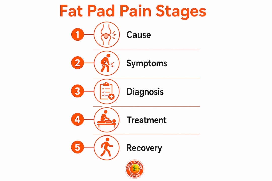
The infrapatellar fat pad, or Hoffa’s fat pad, cushions the knee and contributes to joint lubrication. Inflammation from trauma, overuse, or biomechanics causes sharp anterior knee pain and can lead to fibrosis if untreated. Early diagnosis with clinical exams and imaging guides conservative treatment, including physiotherapy, activity modification, and injections to prevent progression.
The knee fat pad, clinically known as Hoffa’s fat pad or the infrapatellar fat pad, is a specialised cushion of adipose tissue sitting directly beneath the kneecap, inside the knee joint. It is one of the most richly innervated structures in the knee, which explains why inflammation here produces such sharp, localised pain. Knee fat pad syndrome is an increasingly recognised cause of anterior knee pain, yet it remains underdiagnosed because its symptoms overlap with other common knee conditions. Understanding what this structure does, why it becomes irritated, and how to treat it effectively is the first step toward lasting relief.
What is the knee fat pad and what does it do?
The infrapatellar fat pad occupies the space between the patellar tendon, the lower edge of the kneecap, and the front of the tibia. Its primary role is mechanical: it absorbs compressive forces during weight-bearing, reduces friction between moving structures, and helps distribute load across the joint. Because it contains a dense network of nerve endings and blood vessels, even minor irritation produces a pain response that feels disproportionately intense compared with the size of the tissue involved.
The fat pad also plays a secondary role in joint lubrication. It contributes to the production and distribution of synovial fluid, which keeps the joint surfaces moving smoothly. When the fat pad becomes swollen or fibrotic, this function is compromised, and the entire knee mechanics can shift. Patients often describe a deep ache at the front of the knee that worsens when they straighten the leg fully or stand for extended periods.
Fat pad inflammation can also affect patients with knee osteoarthritis, where inflammatory processes within the fat pad contribute directly to pain severity and disability. This makes the fat pad a clinically significant target, not just a passive bystander in knee pain.
What causes knee fat pad inflammation and injury?
Knee fat pad inflammation arises from several distinct mechanisms, and identifying the correct one shapes the entire treatment approach. Direct trauma to the front of the knee is the most obvious cause, but repetitive microtrauma is far more common in clinical practice. Each small compression triggers a low-grade inflammatory response, and over time this leads to hypertrophy and fibrosis, creating a vicious cycle that becomes self-perpetuating without intervention.
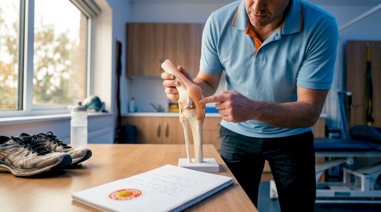
Hyperextension of the knee is a particularly damaging pattern. When the knee locks back into full extension, the fat pad gets pinched between the femur and tibia. Athletes in sports requiring repeated knee straightening, such as gymnastics, volleyball, and distance running, are especially vulnerable. Abnormal biomechanics at the hip, knee, or foot also increase the load placed on the fat pad, even during ordinary daily activities.
Standing with hyperextended knees increases fat pad compression and can worsen symptoms significantly. Many patients are unaware they habitually lock their knees when standing, making postural correction a non-negotiable part of management.
Common contributing factors include:
Repetitive knee extension activities (running, jumping, cycling)
Direct blows to the front of the knee
Habitual knee hyperextension when standing
Patellofemoral pain syndrome or osteoarthritis
Poor hip and foot alignment increasing medial or lateral load
Sudden increases in training volume or intensity
Previous knee surgery altering joint mechanics
Pro Tip: Check your standing posture right now. If your knees are pushed back rather than softly bent, you are compressing your fat pad with every minute you stand. A slight micro-bend in the knee immediately reduces that pressure.
How is knee fat pad syndrome diagnosed?
Accurate diagnosis of fat pad syndrome requires a combination of clinical examination and imaging, because inflammation severity does not correlate with systemic blood markers, making blood tests unhelpful. The hallmark symptom is sharp anterior knee pain that worsens with full knee extension, particularly when the leg is loaded. Patients frequently report pain when rising from a chair, walking downstairs, or standing after prolonged sitting.
The Hoffa test is the primary clinical assessment tool. The clinician applies pressure to either side of the patellar tendon while the patient extends the knee. Reproduction of the patient’s familiar pain constitutes a positive result. Physical examination findings also include tenderness directly over the fat pad and pain reproduced with quadriceps contraction. A skilled clinician can often diagnose fat pad syndrome from examination alone, but imaging adds important detail.
Pro Tip: Tell your clinician exactly when and where your pain is sharpest. Anterior knee pain that peaks at full extension, rather than mid-range, is a strong pointer toward fat pad involvement rather than patellofemoral or meniscal pathology.
The table below compares the two main imaging methods used in diagnosis:
Imaging method | Best used for | Key advantage | Limitation |
MRI | Acute fat pad oedema and structural changes | Highest sensitivity for acute inflammation | Less reliable in chronic phase |
Ultrasound | Dynamic impingement assessment | Real-time movement visualisation | Operator-dependent accuracy |
MRI and ultrasound findings are more reliable during the acute phase of inflammation, when oedema produces clear signal changes. In chronic cases, imaging may appear relatively normal even when symptoms are significant, which is why clinical correlation always takes priority over imaging alone.
What treatment options are available for knee fat pad injuries?
Conservative management is the first-line treatment for the vast majority of fat pad injuries, and it works well when applied consistently. Standard conservative care begins with one to two weeks of activity modification, followed by a structured programme of strength and mobility exercises. The goal is to reduce compressive load on the fat pad while rebuilding the muscular control that prevents future impingement.
A typical conservative rehabilitation plan follows these steps:
Activity modification. Reduce or temporarily stop activities that provoke pain, particularly those involving repeated knee extension under load.
Pain and swelling control. Apply ice for 15–20 minutes after activity. Patellar taping techniques can offload the fat pad and provide immediate pain relief.
Quadriceps strengthening. Begin with isometric quadriceps contractions in a pain-free range, progressing to partial squats and leg press as symptoms settle.
Hip and calf strengthening. Weak hip abductors and calf muscles alter knee alignment and increase fat pad stress. Targeted exercises for these areas are non-negotiable.
Movement retraining. Correct habitual hyperextension through gait retraining and postural education, both standing and during sport-specific movements.
Gradual return to activity. Use a graded loading programme, increasing demand by no more than 10% per week, to avoid re-irritation.
For patients who do not respond to conservative care, ultrasound-guided corticosteroid injections are the next step. Injections reduce pain by 75% in non-osteoarthritis patients and improve function by 52% in osteoarthritis patients within six months. That is a clinically meaningful result, and it supports injections as a legitimate bridge between conservative care and surgery. Ultrasound guidance ensures the medication reaches the correct location within the fat pad rather than the surrounding tissue.
Surgery is reserved for refractory cases where conservative and injection-based treatments have both failed. Arthroscopic resection of the fat pad can relieve chronic impingement, but it carries the risk of reducing the joint’s natural cushioning. Chronic fat pad impingement can progress to fibrosis or osteochondroma if left untreated, which underlines why early, consistent treatment matters far more than waiting to see if symptoms resolve on their own.
Prevention of recurrence rests on three pillars:
Maintaining quadriceps and hip strength year-round
Avoiding habitual knee hyperextension in all daily activities
Progressing training loads gradually rather than in sudden spikes
Pro Tip: Patellar taping, applied by a physiotherapist, can reduce fat pad compression during the early rehabilitation phase. Ask your clinician about McConnell taping technique, which lifts the patella slightly and takes direct pressure off the fat pad.
How can physiotherapy help manage fat pad pain?
Physiotherapy is the cornerstone of fat pad syndrome management because it addresses the root causes rather than just the symptoms. A physiotherapist begins with a full biomechanical assessment, examining how you walk, how your hip and foot alignment affects knee loading, and where your strength deficits lie. This assessment determines which specific tissues are driving the problem and shapes the entire treatment plan.
Localised mechanical stress on the lateral portion of the fat pad causes stiffness and impingement, which means treatment must target specific areas rather than applying a generic knee programme. Physiotherapy techniques used in fat pad rehabilitation include:
Patellar taping to offload the fat pad during early recovery
Soft tissue work to reduce swelling and improve tissue mobility around the joint
Neuromuscular retraining to correct movement patterns that cause repeated impingement
Progressive loading of the quadriceps, hip abductors, and calf muscles
Gait retraining to eliminate hyperextension during walking and running
Recovery timelines vary depending on how long symptoms have been present. Acute cases typically respond within four to six weeks of consistent physiotherapy. Chronic cases, where fibrotic changes have already developed, may require three to six months of structured rehabilitation. Understanding rehabilitation protocols from the outset helps patients set realistic expectations and stay committed to the process.
The physiotherapist also plays a critical role in patient education. Many patients with fat pad syndrome continue activities that perpetuate the problem because they do not realise the connection between their movement habits and their pain. Clear, specific guidance on managing knee pain at home, combined with clinic-based treatment, produces significantly better outcomes than either approach alone.
Key takeaways
The infrapatellar fat pad is a modifiable pain source that responds well to early, targeted treatment combining movement correction, strengthening, and, where needed, ultrasound-guided injection.
Point | Details |
Early diagnosis matters | Chronic impingement can lead to fibrosis or osteochondroma, making prompt treatment critical. |
Hyperextension is a key driver | Habitual knee locking compresses the fat pad and must be corrected as part of any treatment plan. |
Imaging has limitations | MRI and ultrasound are most reliable in the acute phase; clinical examination remains the diagnostic anchor. |
Injections show strong results | Ultrasound-guided corticosteroid injections reduce pain by 75% in non-osteoarthritis patients within six months. |
Physiotherapy addresses root causes | Strength deficits, gait faults, and postural habits all require correction to prevent recurrence. |
A clinician’s view on fat pad injuries
Fat pad syndrome is one of those conditions that gets missed far too often, and the consequences of a delayed diagnosis are real. I have seen patients who spent months treating what they thought was patellofemoral pain, only to discover the fat pad was the primary driver all along. The distinction matters because the treatment approach differs in important ways, particularly around how aggressively you load the quadriceps in the early stages.
The single biggest mistake I see patients make is continuing to stand and walk with hyperextended knees throughout their rehabilitation. They do their exercises diligently, but then spend eight hours a day compressing the very structure they are trying to heal. Postural correction is not optional. It is the foundation on which everything else is built.
Dynamic assessment is also underused. Watching a patient walk, squat, and perform a single-leg stance tells you far more about fat pad loading than a static examination ever will. The lateral portion of the fat pad is particularly vulnerable to stiffness and impingement, and you can often identify the problem pattern within the first minute of watching someone move.
My strongest advice to anyone with persistent anterior knee pain is this: do not wait. The longer fat pad inflammation continues without proper management, the greater the risk of structural changes that are much harder to reverse. A thorough assessment from an experienced physiotherapist, combined with clear guidance on movement habits, gives you the best possible chance of a full recovery without surgery.
— Ivan
Knee fat pad treatment at Parkstherapycentre
Parkstherapycentre has been providing specialist musculoskeletal care since 1986, with clinics across Bedfordshire and Buckinghamshire. If you are experiencing persistent anterior knee pain, a thorough assessment from one of our physiotherapists can identify whether the infrapatellar fat pad is involved and map out the most effective path to recovery.

Our team offers physiotherapy for knee pain alongside biomechanical assessment, patellar taping, and exercise rehabilitation tailored to your specific presentation. For patients who need further intervention, we work alongside clinicians who provide ultrasound-guided injections. Book an assessment at Parkstherapycentre and get a clear diagnosis, a structured plan, and the support to follow it through.
FAQ
What is Hoffa’s fat pad?
Hoffa’s fat pad is the infrapatellar fat pad, a cushion of adipose tissue sitting beneath the kneecap inside the knee joint. It absorbs compressive forces and reduces friction between the patellar tendon and the joint surfaces.
How do I know if my knee pain is from the fat pad?
Sharp anterior knee pain that worsens at full knee extension, particularly when rising from a chair or standing for long periods, is a strong indicator. A clinician can confirm this using the Hoffa test during physical examination.
Can fat pad syndrome heal without surgery?
The majority of fat pad syndrome cases resolve with conservative management, including activity modification, physiotherapy, and patellar taping. Ultrasound-guided corticosteroid injections are effective for cases that do not respond to physiotherapy alone.
How long does fat pad inflammation take to heal?
Acute cases typically improve within four to six weeks of consistent physiotherapy. Chronic cases with fibrotic changes may require three to six months of structured rehabilitation before symptoms fully resolve.
Does knee osteoarthritis affect the fat pad?
Patients with knee osteoarthritis frequently show infrapatellar fat pad inflammation that contributes to pain and disability. Targeting the fat pad directly in these patients can improve outcomes beyond standard osteoarthritis management.
Recommended